Vision is so important to humans that almost half of your brain’s capacity is dedicated to visual perception.
What are the structures of the eye?
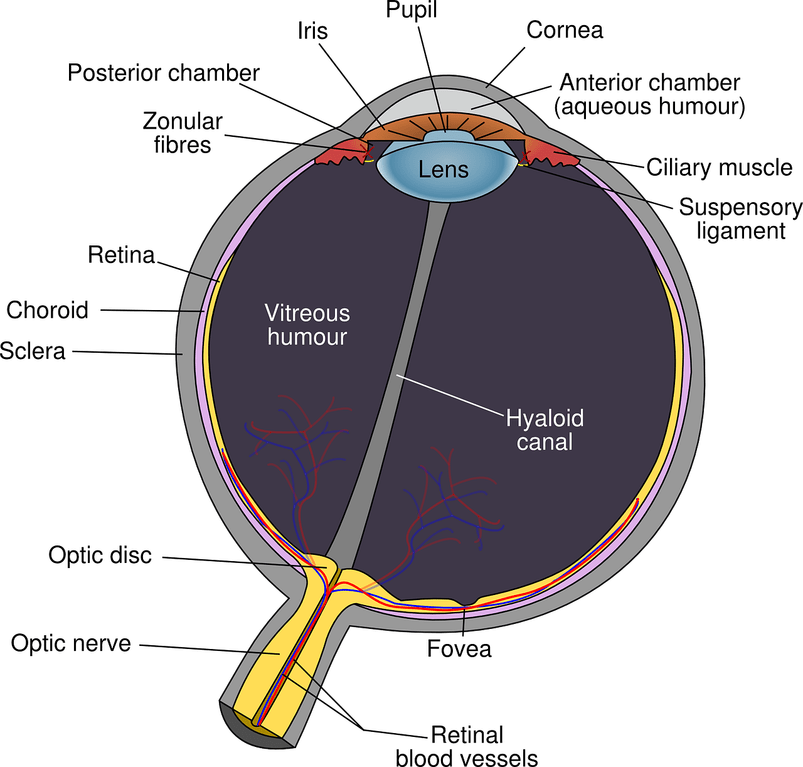
If you visit an ophthalmologist, you may hear some lingo that is not immediately familiar. Here is a basic guide to the important structures of the eye:
- Anterior chamber: Between the iris and the cornea, this space is filled with fluid (the “aqueous”) that nourishes the front structures of the eye. Ophthalmologists often search this location for blood, inflammatory cells, or infectious material that shouldn’t be there!
- Conjunctiva (not labeled): This is the clear, skin-like layer that starts at the edge of the cornea and covers the outside of the eye. It is meant to remain moist with the lubrication of your natural tears. When we are exposed to irritants or don’t sleep well, the arteries and veins of this structure may expand and make our eyes look red. Conjunctivitis, commonly referred to as “pink eye,” also occurs in this location.
- Cornea: This is the clear, curved tissue at the front of the eye. When you wear a contact lens, it sits on the surface of the cornea. This is one of the structures responsible for focusing a clear image onto the retina. Injuries, infections, or dryness of the cornea can be very painful because it is home to many sensitive nerves.
- Fovea: The fovea is a tiny area of the retina that is most critical for good central vision. The fovea is positioned in the center of the macula. Medical conditions that affect this precise area can be quite significant to the vision!
- Iris: The iris is the pigmented (a.k.a. colored) tissue that sits inside the front part of the eye. This is what gives people the appearance of brown or blue eyes. The iris is a muscular structure that opens and closes to allow the appropriate amount of light into the eye.
- Lens: Shaped like an M&M candy, the lens is a structure that sits in the front part of the eye just behind the iris. This provides much of the focusing power of the eye. In childhood, the lens is nearly perfectly clear. As we age, the lens starts to darken to shades of yellow/brown and become much more cloudy. This is called cataract formation. Eventually, when glasses cannot sufficiently sharpen the vision through a hazy cataract, surgery is often recommended.
- Macula (not labeled): This is the central area of the retina that captures the images we see when we look straight ahead. Portions of the retina that are not in the macula are more important for our peripheral vision.
- Pupil: Not exactly a structure in itself, the pupil is actually just the hole that exists in the center of the tissue of the iris. We administer dilating drops during your eye exam to make the pupil as big as possible. This allows the best possible examination of the structures in the back of the eye.
- Optic Nerve: This structure can be thought of as the data cable that connects the eyeball to the brain. When an image hits the retina, the corresponding information is funneled through the optic nerve to the appropriate portion of the brain. The optic nerve may be affected by the pressure of fluid inside the eye. If vision is lost because the optic nerve was damaged by eye pressure, this is commonly referred to as glaucoma.
- Retina: If the eye were to be compared to a camera, the retina is the film that perceives light and begins to form an image. The retina lines the inside of the eye like wallpaper that covers nearly its entire back surface. If the retina is disturbed from its normal anatomical position, such as in the event of a retinal detachment, the impact on vision can be quite severe. Additionally, conditions such as diabetes or macular degeneration may cause blood or other fluids to collect in or around the retina.
- Sclera: This is the white tissue that maintains the shape of the eye. Occasionally, it is the site of inflammatory eye conditions.
- Vitreous: Similar in consistency to egg whites, the vitreous is a gel that fills the large compartment in the back of the eye. As we age, the vitreous tends gradually liquefy. When entering light hits these looser bits of vitreous, tiny shadows are cast on the retina. We often perceive these as floaters that may come and go as we move our eyes.
- Zonules: These fragile cables support the lens in its natural position in the eye like a series of scaffolds. The zonules provide critical stability to the eye when we perform cataract surgery. Zonules may be damaged after head trauma or as a result of certain medical conditions.
Why do I need glasses?
If you’re like me, the world is quite blurry before putting on a pair of glasses or contact lenses in the morning. Why is this?
In a perfect world, all of us would have eyes shaped nearly like perfect spheres. When looking at an object, the light it gives off would hit the front of our eyes and be focused perfectly to a single spot on each retina. This would give us a sharp image with good depth perception as our two eyes work together.
In the real world, not all of us have perfectly shaped eyes! When our eyes can’t provide us with crystal clear vision without glasses or contacts, the term we use is “refractive error.” Refractive error can also be treated with LASIK or other forms of laser vision correction.
What is near-sightedness?
Myopia
The most common type of refractive error is “myopia,” or near-sightedness. You probably have myopia if you can see up close but struggle to see across the room without glasses.
Myopia usually occurs for a couple of reasons:
- If the eye is longer than “normal,” the focusing parts of the eye will create an image in front of the retina. This usually results as the eye grows from its child to adult size. Sometimes, it is hereditary.
- High myopes beware! If the eye gets too long, the risk of retinal tears or detachments increases significantly. This occurs because the retina is stretched along the entire contour of a long, myopic eye. Generally, I advise patients with a glasses prescription of -6.00 or stronger to avoid contact sports and be cautious of new symptoms including floaters, flashes of light, or a shade in the vision.
- If the eye’s focusing parts are stronger than usual, this will also lead images to be created in front of the retina. This might happen in the following examples:
- advancing cataract
- swings in the blood sugar related to diabetes
- pregnancy
Usually, myopia is corrected simply with a pair of minus-powered glasses or contact lenses that move the image backwards onto the retina. Myopia tends to be a benign condition that is easily treated.
What is far- sightedness?
Hyperopia
Hyperopia refers to another common condition where a person’s eyes see well for distance but struggle to focus on objects that are near. The common term for this is “far-sightedness.”
Usually, hyperopia occurs when the eye is shorter or smaller than “normal.” This causes the focusing parts of the eye to place an image behind the retina. We generally treat this with plus-powered glasses or contact lenses that move the image forward.
You may recall that plus-powered glasses are often sold at the drug store in the form of OTC readers. Some hyperopes are lucky – if your prescription is mild and similar between the two eyes, some people can use OTC readers to improve their distance vision instead of prescription glasses. In the long run, this can save a lot of money!
High hyperopes beware! As the eye gets smaller and shorter, its different anatomical structures must fit into a tighter space. This means that far-sighted patients may be at higher risk of an eye emergency called acute angle closure. In this condition, the eye’s own structures may block the “filters” that typically allow fluid to exit the eye. As a result, the pressure inside the eye may rise significantly – even to double or triple what is normal. The symptoms of acute angle closure include severe eye pain, decreased vision, redness, haziness of the front part of the eye, and change in the size of the pupil. If you suspect this condition, visit your ophthalmologist or local emergency room immediately!
What is astigmatism?
Astigmatism
The final common cause of refractive error is astigmatism. This is often combined with myopia or hyperopia. Astigmatism causes multiple images to be formed on or around the retina. As you might have guessed, this can cause vision to be blurry at both distance and near.
Astigmatism occurs when the eye’s contour is curved more in one direction than the other. A common way to think about astigmatism is to consider the difference between a basketball and a football:
- A basketball is a perfect sphere. Therefore, it is equally curved in all directions.
- A football, on the other hand, is more curved in one direction than the other.
When a person has astigmatism, the eyes are generally shaped more like footballs than basketballs. This causes the uncorrected vision to be blurred.
Fortunately, simple astigmatism is also usually easy to correct with glasses or contact lenses. When contact lenses are prescribed to correct astigmatism, the word we use is “toric.” There are even toric intraocular lenses that can be used to correct astigmatism during cataract surgery!
Sometimes, astigmatism is not so simple. This can occur after eye injuries or because of other medical conditions. Your ophthalmologist can help explain your options if this pertains to you.
What are the structures of the eye?


